Sunday, May 8, 2011

"Intelligence Alone is Insufficient"

Moving to New York was the best decision in my life.
I have fond memories of myself as once-perpetually-lonely Floridian anonymously roaming the NYC streets; losing my thoughts happily to day-dream snapshots of other peoples lives while also gaining calm confidence in my independence. Admittedly, this hardening city did take its toll on me at times (The aggressive noisy crowds, miles of monstrous architecture, millions of people 'better apt' for your dreams, and heartbreak) but it functioned to solidify myself in myself. I've spent five years yearning that my future be the present but in four short months I'll be on the road to obtaining my Ph.D.
I must admit I have cold feet.
My love of science and research is probably the only thing in my life that was derived solely from an un-mappable original spot in my body -not from the combination of my parents genes or my particular combination of gene's activity from nurture . (Don't get me wrong, I wanted to do everything and my parents fostered every whim completely). I don't know if anyone, sharing similar genes or not, believed I was going to be a scientist so I kept quiet.
Well, that is, until I came to Columbia University in 2008 and now I will not shut up.


Today's paper: The bHLH factor Olig3 coordinates the specification of dorsal neurons in the spinal cord from/by Thomas Müller1,3, Katrin Anlag2,3, Hendrik Wildner1, Stefan Britsch1, Mathias Treier2,4, and Carmen Birchmeier1,5.


Will not be discussed...Yet!
I have made myself ridiculously busy with my own project, planning grad school, applying for random opportunities, etc.etc...I've also come to realize I need a break from time to time. Anyways, I plan to be back to my normal science blabber soon but for now you can read my first draft of an abstract for our department's retreat.


FATE MAPPING OF DEVELOPING SPINAL SOMATOSENSORY NEURONS


The somatosensory system mediates the perception of touch, temperature, pain, vision and limb position and is essential for sensory regulation of movement. Information from the periphery is transmitted, via primary sensory afferents, to the dorsal horn of the spinal cord where second order sensory projection interneurons relay information via ascending tracts to nuclei in the brain and to other spinal locales. Although much is known about the mature anatomical and physiological organization of the spinal somatosensory system it remains unclear how these complex circuits develop. During mammalian embryogenesis, dorsal spinal neurons arise in the dorsal neural tube from a precursor pool exposed to spatially and temporally restricted signaling cues. From three exclusive bHLH-expressing progenitor domains (Math1, Ngn1, and Mash1) six classes of dorsal interneurons (dI1-6) are produced and are distinguished from each other through differential expression of transcription factors. dIs migrate within the developing dorsal horn and extend axons into contralateral or ipsilateral spinal tracts and eventually populate the entire dorsal horn. To understand the links between early neuronal identity and the subsequent neuronal positioning, axonal trajectory and acquisition of somatosensory modality we are studying the development of one subset, the Math1 derived neuronal population (dI1), using a combination of standard and inducible transgenic mouse lines. dI1 neurons differentiate closest to the roof plate, express Lhx2 and Lhx9 (LIM-HD transcription factors), and migrate to deep dorsal horn where they extend axons either contralaterally or ipsilaterally. The choice to project contralaterally or ipsilaterally is a fundamental decision faced by all projection neurons in the CNS. Studies to date have focused on the regulation of commissural neuron development and there is little information regarding the genesis and function of the ipsilateral population. To understand the basis of ipsilateral character and choice we have generated conditional reporter mouse lines in which dI1 populations can be selectively and permanently labeled at distinct developmental time points. We are using this fate mapping system in conjunction with peri/postnatal retrograde labeling of brain regions to explore the mechanisms underlying the segregation of the ipsilaterally projecting (dI1i) population and their functional importance to the adult sensory pathways. Characterization of these processes may result in novel targets and/or designs to aid regenerative therapies following stroke or spinal cord injury.

Thursday, January 6, 2011

Happy New Year

I work on the 11th floor in a building 22 stories high designed with no 13th floor. It is casually referred to as the Black Building although it is tan in color. The building is named after William Black. Until this entry I had never wondered who he was. I assumed he was one of the many MD alumni to give a large endowment. On the contrary, he is the founder of Chock full o'Nuts whose best friend had developed Parkinson's Disease. Black became the first American to start a private foundation funding scientific research for a specific disease and gave tons of money to Columbia to make a building intended to house Laboratories. There are four elevators but one is a freight elevator only to be used by the custodial staff and people carrying animals. Usually only half of the Otis-made vertical people movers are functioning or take more than ten minutes to arrive. So I take the stairs. I thought about William as I passed the 7th floor today - I wonder if he has even been in the building...

As a young scientist, especially at Columbia, I am taught (and heard the complaints from Grad students) that one should be going to every seminar, doing back-to-back experiments, refreshing pubmed search every hour, and reading papers in every spare minute. Those who succeed are consumed with this process and sometimes smell bad.
While I am consumed by this lifestyle and am ready to throw myself completely into a project - I am scared. If I throw myself into my work completely I'm afraid I'll lose everything else about myself. I want it all. I don't want to be like everyone else and I don't think I have to be. I just wish people didn't judge you for not filling and/or following the footprints perfectly.

Science: I've just finished reading Motor and Dorsal Root Ganglion Axons Serve as Choice Points for the Ipsilateral Turning of dI3 Axons by Oshri Avraham,1 Yoav Hadas,1 Lilach Vald,1 Seulgi Hong,2 Mi-Ryoung Song,2 and Avihu Klar1 published in the Journal of Neuroscience. This entry will discuss the study.


This image depicts the six classes of dorsal interneurons to arise during development in the spinal cord (Embryonic days E9.5 to E13.s in mice) adjacent to the roof plate. From the roof plate a gradient mainly of BMPs and Wnts influence newly born cells based on time of birth and position to express a specific neural progenitor gene. Each of the six classes are produced by a unique neural progenitor and can be identified by a unique combination of known transcription factors, their settling pattern and axon trajectory. In my lab we mainly focus on the D1 class of interneurons (and I primarily am focused on examining further genes/gene expression patterns that which instruct these cells to mature into their less characterized sub populations and eventually use our mouse model to uncover these cells contributions to the adult somatosensory networks).
In this paper, the authors examine the axon trajectory of the D3 interneurons.


Interneuron development/patterning in the spinal cord begins around E9.0 by various gradient produced cocktails of morphogens inducing expression of neuronal progenitors. The progenitors in turn promote the expression of various BarhL and LimHD proteins that not only help identify specific neuronal types but also seem to direct axonal trajectory based on their levels of expression in the cell (Wilson et al. 2008)(However, little is known regarding the targets and/or the mechanisms by which these proteins function).
The path of axon projections is said to be governed by the growth cone interactions with various immediate cues (guiding them to turn their terminals in different directions) and long range cues (repulsion from the roof plate/attraction by the floor plate/later repulsion by the floor plate after crossing). This paper proposes that at least one group, d3, uses architecture and possibly, Isl1 (cell-cell interaction), to guide them in their turning decisions when projecting laterally out from the spinal cord to their final targets.


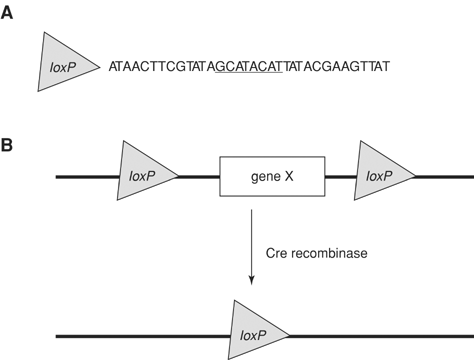
First the authors sought to characterize the population of cells in more detail. We know that these cells express Tlx3,Prrxl1, Brn3a, Olig3, Asc1 and they are the only interneurons to express Isl1 (which is also expressed in Motor and Dorsal Root Neurons). They searched for evolutionary conserved non-coding enhancer elements around the Isl1 gene via Enhancer Browser Project. They cloned three different regions fused CRE recombinase downstream of the enhancer and screened for labeling by electroplating Chick neural tubes along with a conditional nGFP reporter vector. They found a combination of using two elements specially labeled the dI3 cells (referred to as: EdI3). They subsequently used this system to label and observe the intrinsic nature of the cells.

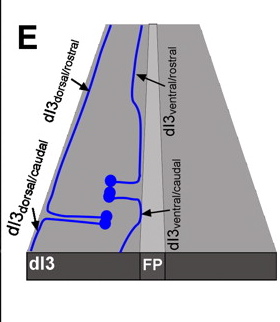
Based on axon projection and turning point for lateral growth they defined four distinct populations of DI3 neurons: Dorsal projecting turning caudal or rostral at dorsal funiculus, Ventral projecting turning caudal or rostral at ventral lateral funiculus. The ventral projecting axons tend to turn/follow the motor neuron axons out of the ventral spinal cord and the dorsal projecting neurons follow the DRG neurons out of the dorsal cord to then turn laterally towards final targets.
The top figure above illustrates the four types of Di3 neurons identified as observed in 'open book presentation'. Basically they took the roundish spinal cord and cut down the midline from roof plate to the floor plate and opened the cord like a book. See illustrated example below from Dickson BJ, Gilestro GF (2006) Regulation of commissural axon pathfinding by slit and its robo receptors. Annu Rev Cell Dev Biol 22: 651–675.


The authors further state that the axons of the dorsal interneuron classes do not intermingle, their cell soma positions get more mixed between each class as they migrate ventral or dorsal but their longitudinally projecting axons bundle together specific to their class.

They go on to investigate the molecular actions of Isl1. The LimHD genes have previously been shown to play no role in cell fate (Avraham et al., 2010; Kania and Jessell, 2003; Pillai et al., 2007; Luria et al., 2008; Wilson et al., 2008) but they are emerging to have roles in maintaining a cells fate and their axon patterns. Avraham electroporated Chick neural tubes at HH14 (premitotic) and HH19 (postmitotic) with nGFP and Lhx9 or Isl1 then quantified the protein expression of the limHD proteins compared to the control (un-electroporated side). They found that there is a cross-repressive relationship between Lhx9 and Isl1 in Pre- and Postmitotic cells. Aberrant expression of Lhx9 imparts a reduction in Isl1 protein as compared to the controls. A similar decrease in Lhx9 protein is observed when electroporated with Isl1. They also tested and found cross-repression between Lhx1 and Isl1.

The expression patterns of other transcription factors known to mark these cells are not changed (Brn3a, Pax2, Tlx3). Avraham concludes that ectopic expression of Isl1 does not affect the fate of other cells and does not induce a population increase of dI3 neurons. He supports his conclusion by taking a look Isl1hypo- mutant mouse. In this mouse line the motor neurons express some Isl1 but the dI3 neurons are lacking Isl1 protein completely. So the dI3 cells are generated even in the absence of Isl1 - they just must not project correctly.

Next they directed ectopic expression of Isl1 in dI1 class neurons in the Chick cord (they used the specific enhancer element expressed in dI1 neurons with CRE fused downstream and electroporated the construct with a Lox'd Isl1 taumyc tagged expression construct) to examine if expression of the Isl1 gene would confer a dI3-like axonal projection pattern on the dI1 neurons.
The dI1 neurons send axons ventrally towards the floor plate and cross the midline turning in the ventral lateral funiculus (contralateral/commissural) but they also send axons ipsilaterally (same side) in the medial towards ventral lateral funiculus. While some dI3 neurons project dorsally none of the dI1 neurons send their axons in the dorsal direction. The authors state about 20% of dI1 neurons project to the contralateral side but in the dI1-Isl1 expressing neurons only 10% cross the midline. They observed these neurons to turn laterally upon reaching the motor neuron zone, follow their tracks (like dI3-ventral neurons) and turn laterally. They found no change in the dI1-ipsilateral-Isl1 expressing neurons.


Is this due to timing of expression, Higher Lhx9 expression in the ipsilateral population (Wilson 2008) allowing for functional repression of Isl1, or (as the Authors discuss) the axons of the dI1-ipsi never get close enough to the motor neurons?

They discuss that Isl1 may infer a short range adhesive interaction between the motor neurons and dI3 neurons that is required for their turning lateral. They do state that this can be challenged by manipulating the motor neurons/DRG neurons.
I think they should have also targeted Isl1 reduction in motor neurons. If you knock down Isl1 in the motor neurons - do the ventral projecting dI3 neurons or dI1-contra-isl1 expressing neurons still turn laterally upon reaching them?

Although LimHD protein repression was illustrated as a molecular consequence of Isl1 expression, I wonder what the other targets of Isl1 are (and the other LimHD proteins specific to the other dorsal classes). There must be other players in the scheme that are repressed or promoted....

Tuesday, December 7, 2010

Of interest

Hello, It has been awhile Internet world!

After I finished my applications for admission to graduate school I jumped on a plane to Europe. I celebrated thanksgiving in Sweden. It was magical - Stockholm seemed like a beautiful snow globe and the people were just as amazing. I did stop in London and Paris, both cities are great as well, but for some reason I have a strong connection with Sweden.

Regardless, I'm back in lab with tons to do.
Today I received a new batch of Math1-Lacz spinal cords back-filled from the thalamus with Fluorogold and last week I prep a huge litter of E19.5 Tamox-induced (@E8.5) Math1-myrGFP spinal cords for examination.
I am really looking forward to examining these cords and getting an idea of what’s going on. Especially the Tamox line - I haven't looked at spinal cords older than E13.5 Sooo it will be interesting to see if I can detect any projections into the brain at this stage. (Or really detect anything at all - the literature and in situ's done suggest that Math1 expression in the spinal cord begins at E9.5 and continues till E13ish. Therefore, my tamoxifen treatment may not have induced any GFP expression OR dependent on how long the tamoxifen stays active in the system it may induce and label the first round of cells to differentiate into the D1 class.)


Anyways, here's your food for thought today.


Please press that link and read the letter.

Monday, November 1, 2010

MTL?!

Soon is the 33rd Annual W. Alden Spencer Award and Lecture is taking place here at Columbia. They are honoring Larry Zipursky and Marc Tessier-Lavigne.
Zipursky intitally discovered and explored the family of Dscam proteins – Oweing to much of our understandings of neuronal circuitry.

Tessier-Lavigne has been a name in my mind for six years now; he has uncovered many mechanisms relating to axon guidance. He has just been donned as Rockefeller University’s new president and has lead quite a wealthy career keeping up with basic research while also pursuing the biotech industry. He is definitely someone I look up to when I imagine life in terms of my own career. I welcome him to New York City in March 2011 (I’m hardly even an ant but still)!

Recently, He published in Journal of Neuroscience with Alexander Jaworski and Hua Long at Genentech on “Collaborative and Specialized Functions of Robo1 and Robo2 in Spinal Commissural Axon Guidance”.
Here are some main discoveries:
They analyzed the amount and ability of axons to cross the midline at the floor plate during project at E11.5 and E12.5 in various genetic backgrounds. Robo1 and Robo2 are the receptors which respond to the Slit1-3 repellants produced in the floor plate.
Robo3.1 is implicated in repressing Robo1’s function therefore diminishing axon repulsion and allowing commissural projecting cells to target their growth towards the floor plate. After midline crossing, Robo3.2 is then unregulated in axons and acts as a classic Slit repellent pushing axon growth towards its next target.
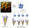
Robo3 mutant animals have a major deficit in commissural projecting neurons in the spinal cord. In previous studies it was found that in the mutant Robo3 background loss of Robo1 partially rescued the deficit, as well as, loss of both Slit1 and Slit2.
In this study they found that in the Robo3 mutant background a loss of both Robo1 and Robo2 restored the crossing deficiency significantly. Indicating the failures of Commissural axons to project through the midline in the Robo3 mutant are partially due to Robo1-depedent repulsion but also Robo1- and Robo2-repulsion.
Due to combination mutants of Robo1/2/3 they end up illustrating that Robo1 and Robo2 work together to prevent post-crossing axons from reentering the midline and they have individualized roles in the sorting of axons into specific positions within the ventrolateral funiculus.
Lastly, I find most interesting, they provide evidence that there is most likely a Robo3-dependent mechanism not involving Robo1 or Robo2 (or the combination of both) allowing midline crossing in a subset of contralateral neurons.


I will post soon on the talks given at the Spencer award & lecture!

Monday, October 25, 2010

Seminar: Embryonic stem cells as a tool to study Motor Neuron development and diseases

Today I attended a PACKED seminar by Columbia's own Hynek Wichterle in the department of Pathology and Cell Biology. The walls were lined with standing people and the floor covered with sitting students.
Hynek's lab studies stem cells in hopes to better understand motor neuron development and diseases.
He began his talk introducing Hans Driesch's experiments with Sea Urchins in 1892. He, studying the embryo, separated the cells resulting after the first round of cell division. He imagined that the cells would then develop into the respective but separated portion of the sea urchin corresponding to the location where the cells were harvested. Instead each of the cells developed into fully functional complete sea urchins.
The initial cells Driesch harvested where embryonic stem cells, totipotent in nature, able to differentiate into any cell (not to be confused with: Pluripotent able to become cells that compose any of three germ layers, multipotent able to become many cells of a related family, Oligopotent differentiate into very few set of cells, and unipotent can only become themselves). So awesome, right? Let's just play with embryonic stem cells. But, really - let's!

In any developed organism the amount of cellular diversity, the systems of cross talk and proximity between these highly diversified cells makes studying specific cells very difficult. In vivo animal mutant models can be tricky and lead to embryonic lethality before gaining any new insight. Therefore, learning more about the genetic pathways and environments needed to induce stem cells to become specific types of cells can allow researchers to study those very cells more concretely. Wichterle aim's to do just that with motor neurons.

Hynek continues on to point out that due to the diligence of labs previous research (please visualize the head nod that ensued in the direction of his previous mentor, Tom Jessell) we know ways to distinguish motor neurons based on molecular identity and functionally. The signals required to develop spinal cord motor neurons are also being elucidated: starting from the rostral or cervical end of the spinal cord (the neck) retinoic acid (RA) signals at its strongest then lessens as a gradient down the spine through the brachial region towards the middle of the cord meeting the least of the increasing gradients of GDF, FGF and WNT (signaling morphogenic molecules).
Wnt, GDF, and FDF increase along the thoracic level till reaching the end of the lumbar or caudal spinal cord. Lastly, across the entire ventral portion of the spinal cord, SHH signals as a gradient strongest from ventral lessening upwards into the dorsal (top half) area of the cord.

Using these tools and signals, the Wichterle lab has been successful in inducing pluripotent stem cells in vitro to differentiate into cervical and brachial motor neurons. They express the appropriate markers, when injected back into a developing spinal cord they migrate to the correct distinct positions, and can then function perfectly as the in vivo developed cells within the tissue. Now with the model cells to work with the lab moves on to test the role of repressive chromatin in the pattern of developing motor neurons in the rostral to caudal axis, microRNA's in the dorsal-ventral axis, and modeling neuronal diseases like ALS.


Hox gene products, homeobox proteins, are an important family of transcription factors during development; they aid in the creation of different motor neuron classes across the Rosto-Cadual axis (length from the neck to tail). Different sets of Hox genes are required to be expressed in different anatomical locales along this axis. For example, starting Rostal, the cervical region requires Hoxc genes 4-5, Brachial requires Hoxc6-8, and (moving caudual) Thoracic Hoxc9 and Lumbar Hoxc10-13.
The hoxc genes are interestingly assembled in the genome on chromosome 12 in a similar fashion to expression; hoxc gene 4-5 are organized in the 3' end in chromatin (DNA) and increase in order of number till the 5' end. Therefore, it is thought there must be repression of specific hox expression in these distinct anatomical locations to allow for the differential gene expression (i.e. repression of hoxc4 & 5 in the lumbar section but none of hoxc10-13 permitting their expression). It was postulated through correlations that during development repression is lifted from the primary hoxc genes, priming the Chromatin structurally, allowing transcriptional molecular machinery access for the later relaxation (de-repression) of the numerically higher hoxc genes. Therefore, it was postulated that there is a progressive clearance of repressors on the chromatin starting with Hoxc4 and continuing to Hox13 during the due time of need for induction of those cells.
Wichterle and Dansen labs collaborated (NYU, NOTE: post-doc from Jessell lab as well) to confirm the postulated nature of the Hoxc chromatin. They performed chromatin immunoprecipitations on stem cells and found that all hox genes had a considerable amount of repressive markers across them. They looked at the different cell types in the cord and found in development cervical motor neurons lack of repressive markers. Puzzling, they decided to do Quantitative PCR to profile the hoxc gene expression during time points in the developmental cascade of inducing stem cells to motor neurons. They found that there was no progressive clearance of repression, the profile of hox gene repression did not correlate with expression. They decided to do more chromatin immunoprecipitations but looked for the density of retinoic acid receptors. They found a correlation between the presence of RA receptors located on the hoxc 4-5 genes and the loss of repressive markers. Wichterle states that somehow the recruitment of RA receptors initiates the clearance of repression. Cool, How?
Its pretty convenient that the two most well know players in the patterning of motor neurons along this axis show tight correlation between their activities. Can you further confirm RA receptors role by stunting it's gradient or prolonging its gradient along the axis, then look at methylation (this is the marker they used for repression, methyl groups added on chromatin usually mark a repressed gene)? That is, at a time when the changing RA gradient won't completely mess up the fate of the cells...?
What are the molecular complexes that are responsible for providing the methylation on the chromatin? What is the gene expression profile for these chromatin remodelers - Micrroarray and look for enrichment of certain known chromatin modifiers?

Wichterle goes on to question how the caudal neurons develop. It seems throughout the development of the cells there is no lack of repressive markers. They looked at a microarray to see what genes were enriched in the lumbar cells versus the cervical. They found one interesting gene, Cdx2. Inducing stem cells with cdx2 seemed to have no role, but they found in cooperation with FGF they could induce expression of hoxc8 motor neurons. Through further experimenting they found they could also induce hoxc6-9 genes but repress hoxc1-4 genes. He concludes showing that hoxc9 likely cooperates in the repression of hoxc1-4. (For more information see paper)
The orchestra is getting meatier regarding the instruments to play out development of even one cell type: Signals, transcription factors and chromatin modifiers...

Sorry for the length of this one...

Hynek continues with part two, what I found to be the most interesting data, about the dorso-ventral patterning of the spinal cord. SHH signals as a gradient from the notocord and floorplate (Ventral/bottom of Spinal cord aka the side facing the front of your body) which is responsible for inducing different varieties of Interneurons and Motor neurons based on the receieved amount of SHH. In the ventral portion, closest to the floor plate, cells are induced to express NKx2.2 as the gradient lessens dorsally the next class of express Olig2 (Motor neurons) and then further lessening of the gradient the next class express Irx3 (picture: http://en.wikipedia.org/wiki/File:Sonichedgehog.jpg). These genes are markers of progenitor cells, the cells expressing them have matured from stem cells and based on gene expression decided to be a specific type of cell. (ex. nkx2.2-motor neurons, olig2-oligodendrocytes).
Hynek's lab investigated the role of microRNAs in cells that border an area with overlapping gradient signals.
Presumably these two gradients are providing the cells with signals that instruct competing progenitor gene expression to either become one type of cell or another. Somehow cells naturally overcome this confusion and the spinal cord results in a clear pattern of segregated neuron types.
The lab noticed previous research done that identified a gene that when knocked out would eliminate all microRNAs, Dicer. Dicer mutants do not live past gastrulation, therefore studying it's affects have been proven difficult. Wichterle's lab formulated a conditional tamoxifen-inducible line for dicer. Allowing them to knockout dicer's expression during specific time frames in development. They still had issues with survival of the animals but had enough success to illustrate an expansion of motor neurons and losses of Irx3 cells. Seemingly, in the natural background microRNA represses the expression of Olig2 allowing only a discrete segment of the spinal cord to develop into motor neurons. This indicates that the p2 set of cells (Irx3 expressing) must transiently express olig2 at some point. The further investigated the situation by looking at the microRNA profile of cells exposed to high vs. low levels of SHH.

To Be Continued...


This is very long. I do apologize. I think I just got really excited about the research...my head is exploding with information.

Friday, October 8, 2010

Seminar: Developing Therapies for motor neuron diseases.

I've been meaning to start this blog for quite some time now. The basic point is that I have too many thoughts; I can end up talking too much and making way too many tangent conversations.
I've created this blog for me to summarize certain seminars I attend, papers I read and just blab about my work/things that interest me. Not only will I be able to clear my mind but I am hoping some will read this and have questions/answer my questions/comment.

Disclaimer:: I do not think I have any literary abilities. Please do not focus on my writing capabilities or grammatical errors.

Here it goes:

Today I attended a talk hosted by the Motor Neuron Center at Columbia University Medical Center. The speaker was Brian Kaspar from Ohio State University, he presented a short talk on his lab's work on Gene therapeutics targeting SMA and ALS.
He began the talk introducing the general problems with targeting diseases in the CNS; the blood brain barrier, diversity of cell types, magnitude of the amount of cells from the Brain reaching down throughout the spinal cord, etc. Kaspar then introduced Adeno-associated viral vectors (AAV). AAV is a virus naturally found in humans and nonhuman primates, it has no known pathological responses and can integrate into post-mitotic cells. Once infected in cells it integrates it's genomic information stably for long term trans gene expression (on Chromosome 19). With these attributes it sounds like an awesome vehicle to use when searching for ways to deliver gene therapies to various live systems. Currently there are clinical trials ongoing for Cystic fibrosis and Parkinson's disease.
The trick is figuring out which family member of the virus to use, how to administer it, when to administer it and what are the effects of the trans gene expression. For example, some familial genetic disorders present themselves because of a lack of a certain gene's expression (therefore, lack of a certain protein). Some gene therapies have a long list of issues in clinical trials because of the body's natural immune response to a protein it has never 'seen' before - even if that protein can rescue the disease over time - one would still need to battle the immune system to allow it. In light of this issue, another problem occurs = lack of focus on efficacy. FDA clinical trials usually focus on the safety of the treatment but not the efficiency to alleviate the disease. Moving on...
The lab initially started testing AAV6-GFP (green fluorescent protein) and AAV8-GFP through tail vein injection but had no success in penetrating the blood brain barrier to the CNS. However, when they tested AAV9 they were very successful in finding GFP expression in a robust amount of astrocytes all over the nervous system and some neurons. Astrocytes are mainly born a few days postnatal. With this in mind they tried to target mostly neurons by injecting intravenously through a facial vein visible postnatal 1 and 2 (tails are too small then). The result was great GFP expression in the dorsal root ganglia and motor neurons throughout the spinal cord and various neurons in the brain (neocortex, hippocampus, cerebellum).
Both scenarios present possible good routes for therapies against either SMA (neonatal injection) and ALS (adult tail injection). SMA is luckily known to be attributed to a loss in the SMN1 gene expression. Therefore, animals affected by SMA have at some point 'seen' smn1 protein physiologically speaking. Using an SMA mouse model, they tested the ability of AVV9-SMN1 treatment to rescue the disease phenotype. In short, they were successful in finding that the treated SMA mice compared to untreated SMA controls were able to become stronger and eventually live normal mouse life spans (tested through muscle growth, behavior, electrophysiology, and life span). However, SMA semi-adult mice (they only live about 15 days) past neonatal do not response similarly to the treatment, as the mice age the AAV starts targeting mainly astrocytes and less neuronal populations which ends up not rescuing the illness.
The Kaspar lab is now testing this gene therapy on non-human primates and having great success. They plan to move forward with human clinical trials.
They are also doing a lot of work on ALS, but I'm tired of writing about it for now. Need to do some image analysis.

Very exciting stuff in terms of possible treatment options.

Here is a link to their recent paper about AAV9 injections: http://www.nature.com/nbt/journal/v27/n1/abs/nbt.1515.html